
If you have a leg diagnosed with a lipoma, or suspect that you can have one, you may wonder about the need for a lipoma exploration.
A lipoma is a benign tumor composed of fat tissue. It is generally soft, mobile and painless. But when it is large, painful or growing, scan may be necessary. It may also be necessary to have a scan if you consider that the lipoma is eliminated.
This article aims to demystify the process of a lipoma exploration. I will explore different types of scanning, such as ultrasound and magnetic resonance, and explain when each one can be used.
I will also discuss what to expect the scan by default, how to interpret the results and possible treatment options. My goal is to provide a clear and empathic guide to help you make informed decisions about your medical care.
Remember, although this article provides general information, it is always better to consult with your doctor to obtain personalized advice. We are going to immerse ourselves and learn more about Lipoma’s scans.
Lipomas understanding: a general letter description
Lipomas are the most common type of soft tissue tumor. These are not channels and consist of fatty tissues. The usual present as soft and gummy packages under the skin.
Most lipomas are painless and do not require treatment. They can appear anywhere in the body where there are fatty cells.
The exact cause of lipomas is not well understood. Genetic factors can play a role in their development.
When a lipoma becomes painful or grows rapidly, medical evaluation becomes important. While they are benign, these changes require attention to rule out other conditions.
Understanding lipomas helps make informed decisions about when more research can be guaranteed, such as scanning.
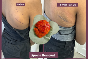
This is a great lipoma in the back. A scan was required before extraction due to its size.
When is a lipoma exploration necessary?
A lipoma exploration becomes vital in specific circumstances. In general, lipomas do not require any intervention. However, certain changes in their behavior can justify a closer look.
If you notice that your lipoma is increasing in size, this may require additional exam. Size changes may indicate the need for more detailed images.
The pain associated with a lipoma can also indicate the need for scan. Althegh, typically painless, some lipomas can press the nerves. There is also a variety or benign lipomas called angiolipomas that can be tender.
In addition, when the diagnosis is not clear from a physical exam, an exploration can help. This is crucial to differentiate a lipoma from other types of growth.
If you are considering that the lipoma is eliminated, a scan can help delineate how deep is your lipoma. This is important to decide white, its lipoma can be eliminated under local or anesthetic anesthesia.
Your doctor could recommend a scan based on certain criteria. Here are some key indicators:
- Rapid lipoma growth.
- Pain or promotion around the area.
- Skin changes on lipoma.
- Difficulty determining the nature of growth: packages around the head and neck can be a variety of things.
- Lipoma location: Lipomas in the trunk can sometimes be deeply in the muscles.
If you experience any of these symptoms, discussing a scan with your doctor is wise.

A swelling in the neck could be a variety of things and a scan would be necessary before elimination.
Types of explained lipoma scans
There are several types of scan available to evaluate a lipoma. Each scan sacrifices benefits and unique levels or details.
Ultrasound is of the first option. It is not invasive and uses sound waves to produce images. This makes it appropriate to examine packages near the skin surface.
A magnetic resonance exploration provides more detailed images. It is special useful for deep lipomas within the body. Magnetic resonances use a magnetic field and radio waves to generate images.
Thought less common, computerized tomographs are another option. They use X -rays to create a detailed cross section of the body. CT explorations are sometimes used for complex cases.
The decision on which the scan is used depends on the specific characteristics of the lipoma. Your doctor will guide you to choose the appropriate scan.
Each type of scan helps to diagnose and plan the next steps. They confirm if a lipoma is harmless or needs more attention.
Understanding the purpose of these scanning can relieve anxiety. Knowing what to expect to make the process feel less discouraging.
Discussing the scans with a doctor will clarify which adapts to their needs. You will have the information to process with confidence.
Lipoma ultrasound exploration
An ultrasound is from the first scanning option to examine lipomas. It is painless and does not require any preparation.
The device uses sound waves to create images inside the body. This helps identify lipomas near the skin.
Ultrasound are effective in distinguishing lipomas from other growth. They provide rapid results and do not imply radiation.
Duration The process, a technician moves a small device on the lipoma. The data is sent to a monitor, producing an image in real time.
This exploration is also safe for those who are pregnant. It is a simple and effective way to examine soft tissue packages.
Lipoma magnetic resonance scan
Magnetic resonance sacrifices a more detailed vision of lipomas than ultrasound. They are ideal for lipomas located in the depths of the body.
MRI use a magnetic field along with radio waves to produce detailed images. They highlight soft tissues and structures around the lipoma.
This type of scan helps determine if a lipoma affects the surrounding tissues. It provides clarity when the edges of a lipoma are not well defined.
Special preparation is not needed for magnetic resonance, and it is without pain. However, patients with metal implants should inform their doctor in advance.
The detailed images of a magnetic resonance aid significantly in the precise diagnosis and treatment planning. It is a superior option when intricate images are needed.
Other scanning techniques
Designed not so common, computerized tomographs are sometimes used for lipomas. They provide transversal images through radiographs.
This method is useful if a magnetic resonance is not adequate for some reason. It is an alternative that provides different image perspectives.
Rarely, other specialized explorations can be considered. Each case is unique, so Doctor’s recommendations are important.
Preparing for your lipoma scan
Preparing for a lipoma exploration is simple and requires little effort. Most scanns, such as ultrasound or magnetic resonance, do not need special preparation.
Your doctor will inform you if there are specific steps to follow before the scan. You can usually eat and drink as usual.
Wear comfortable clothing on the day of the scan and try to relax. This can help you feel more comfortable the duration of the procedure.
What to expect scan duration
When he arrives at his Lipoma Escane, a health professional will guide him through the process. They will ensure that you feel comfortable and informed.
Departing on the type of scan, you can lie on a table or sit in a chair. For ultrasound, a skin gel is applied to better contact.
The scan is generally painless and fast. Ultrasound and magnetic resonance scans generally take between 15 and 45 minutes.
Through the scan, the technician can give instructions. This could include containing breathing briefly to get clearer images.
After scanning, you can resume your normal activities without restrictions. Be sure that professionals are there to support it in each step of the road.
Interpret the results of the exploration with your doctor
Once your scan is completed, your medical care provider will review the images. They will evaluate the characteristics of the lipoma and provide a report.
The report will explain whether the lipoma seems benign or if more steps are needed. It will also give the dimensions of the lipoma and a precise representation of how deep it is.
Do not hesitate to ask any questions about the results. Understanding the details will help you feel safer on the next steps.
The results may guide decisions on treatment or monitoring options. This discussion allows a personalized plan adapted to your needs.
Remember, your doctor is there to support and inform you. Your experience will ensure that you make informed health decisions. If you have any questions about your scanning report, you can always send us a copy and we can help you guide it.
Treatment options after a lipoma exploration
After evaluating your Lipoma exploration, your medical care provider will discuss possible treatment options. Often, if the lipoma is small and harmless, a treatment is not necessary.
However, if the lipoma causes discomfort or aesthetic groups, elimination can be recommended. Surgical split is a common method to eliminate a problematic lipoma. It involves making an incision to extract all growth and something we specialize in the Staian clinic.
We sacrifice a ‘see and treat’ service for the elimination of lipoma where you can send us a photo of your lipoma and a copy of your scanning report (if you have had one). We will give you a price for lipoma extraction and sacrifice a date (usually within 7 days) to come and receive a consultation with one of our plastic surgeons and its lipoma eliminated the same day.
Questions to ask your doctor about Lipoma’s scan
It is important to communicate openly with your doctor about your lipoma scan. To help this dialogue, consider the preparation of a list of questions to clarify any uncertainty.
You may want to ask about the need for scanning and possible results. Understanding the reasons behind your recommended scan can provide tranquility.
Here are some questions that can prospective:
- Why is a scan for my lipoma necessary?
- What kind of exploration would be more appropriate and why?
- Is there any risk or side effects associated with scan?
- How will the results influence my treatment options?
These questions can help guide your conversation. Feel empowered to collect all the information you need to make informed health decisions.
Conclusion: Make an informed decision: Do I need a lipoma exploration?
Your doctor will guide him to make a decision on whether he needs a scan of his lipoma.
There are some characteristics that will make it more likely to need a scan:
- Size – If your lipoma is more than 5 cm in diameter
- Location – If your lipoma is on your neck or your trunk
- Symptoms – If your lipoma is growing rapidly or painful
Choosing whether or not to process with a lipoma exploration can be discouraging. Consult your GP to see if a scan is necessary. If your GP cannot help you, then we can organize a lipoma exploration in one of our partner hospitals that will provide a report that allows us to process with treatment. Call the clinic at 0121-454 3680 for more details.
Ultimately, their comfort and tranquility are more important. The informed elections leads to more effective health decisions. Trust the process and remember that you are not alone on this trip.